Joint Research Group Macromolecular Crystallography
Structure of the month - July 2015
Nature 520 (2015) 571-574.
Hydrogens detected by subatomic resolution protein crystallography in a [NiFe] hydrogenase
Hideaki Ogataa*, Koji Nishikawaa, Wolfgang Lubitza
a Max-Plank Institute for Chemical Energy Conversion, Stiftstrasse 34-36, D-45470 Mülheim an der Ruhr, Germany
* Corresponding author: Hideaki Ogata, Max-Plank Institute for Chemical Energy Conversion, Stiftstrasse 34-36, D-45470 Mülheim an der Ruhr, Germany
E-mail: hideaki.ogata@cec.mpg.de
Phone: +49(0)208-306-3508
Abstract
In protein X-ray crystallography the detection of hydrogen atoms is one of the difficult problems. Because they display only weak contributions to diffraction and the quality of the single crystals obtained is often insufficient to obtain sub-Ångström resolution. The location of the abundant hydrogens is, however, of key importance for understanding protein structure and function. This is particularly true for the enzyme hydrogenase that reversibly converts dihydrogen to protons and electrons at a metal catalyst. We used a BESSY II (HZB, Berlin) to collect a high quality X-ray diffraction data set that was carefully analyzed. Thus, an ultra-high resolution crystal structure of [NiFe] hydrogenase from Desulfovibrio vulgaris Miyazaki F with 0.89 Å resolution was obtained that provides an extraordinarily detailed picture of the enzyme poised in this specific catalytic state (Ni-R) that has not yet been described but is of central importance in the enzymatic cycle. The high resolution, proper refinement strategy and careful modeling allowed the positioning of a large part of the hydrogen atoms in the structure. This has led to the direct detection of the products of the heterolytic splitting of dihydrogen into a hydride (H-) bridging the Ni and Fe and a proton (H+) attached to the sulfur of a cysteine ligand. The Ni-H- and Fe-H- bond lengths were 1.58 Å and 1.78Å, respectively. Furthermore, we could assign the Fe-CO and CN- ligands at the active site, and could obtain the H-bond networks and the preferred proton transfer pathway in the hydrogenase.
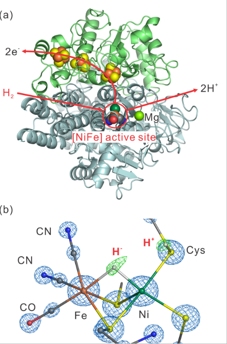
Fig. 1. (a) The overall structure of [NiFe] hydrogenase form Desulfovibrio vulgaris Miyazaki F. The small and large subunits are shown in green and gray, respectively. Schematic representation of the hydrogen, proton and electron pathways is shown with the red arrows. (b) The electron density map of the [NiFe] active site in the Ni-R state. The blue and green meshes represent the 2Fo-Fc and Fo-Fc electron density maps, respectively.
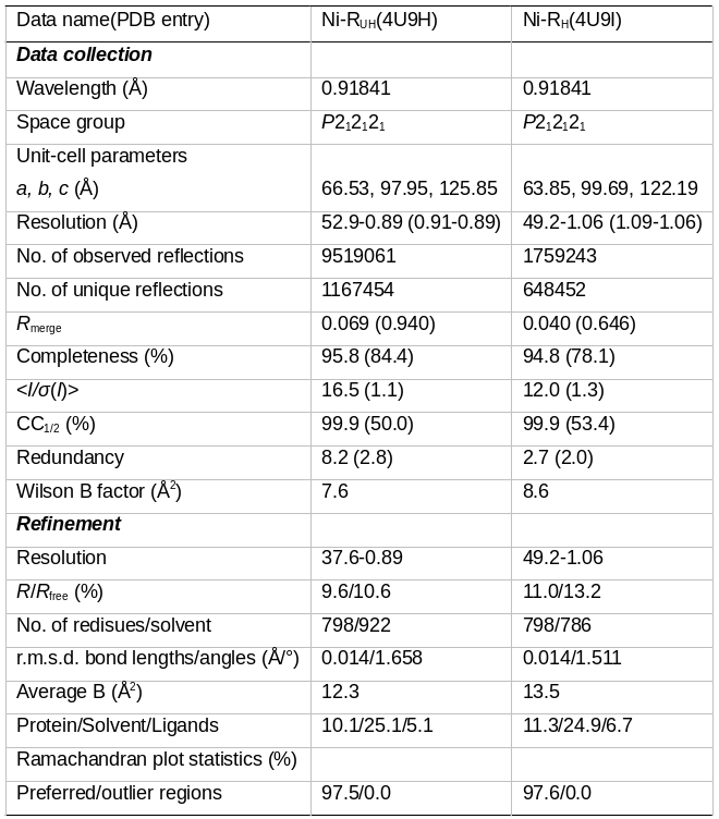
Table 1.