How to Flow Ultrathin Water Layers - A Liquid Flatjet for X-Ray Spectroscopy

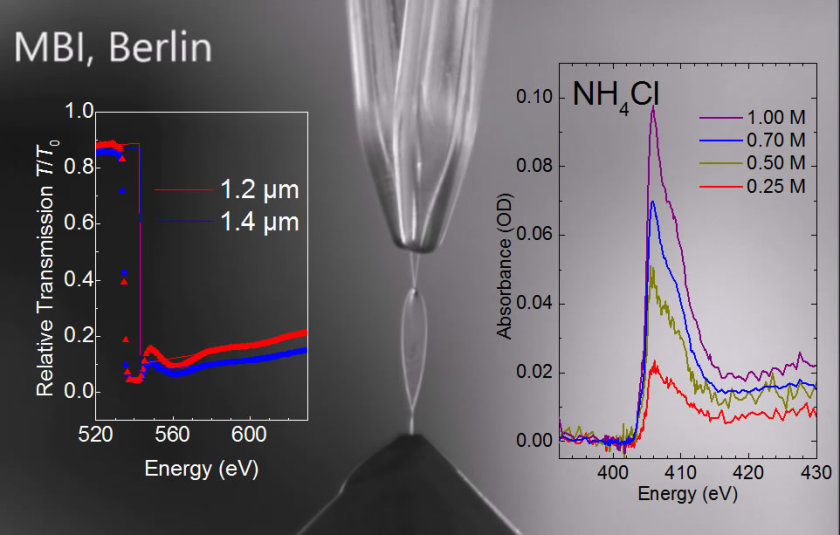
Liquid flatjet system, showing the two nozzles from which two impinging single jets form a liquid water sheet with a thickness of 1 - 2 μm. © MBI
A collaboration between scientists from the Max Born Institute for Nonlinear Optics and Short Pulse Spectroscopy (MBI), the Helmholtz-Zentrum Berlin (HZB) and the Max Planck Institute for Dynamics and Self-Organization (MPIDS) have now demonstrated the successful implementation of a liquid flatjet with a thickness in the μm range, allowing for XAS transmission measurements in the soft-x-ray regime. This paves the way for novel steady-state and time-resolved experiments.
A collaboration between scientists from the Max Born Institute for Nonlinear Optics and Short Pulse Spectroscopy (MBI), the Helmholtz-Zentrum Berlin (HZB) and the Max Planck Institute for Dynamics and Self-Organization (MPIDS) have now demonstrated the successful implementation of a liquid flatjet with a thickness in the μm range, allowing for XAS transmission measurements in the soft-x-ray regime. This paves the way for novel steady-state and time-resolved experiments.
Here a phenomenon well known in the field of fluid dynamics has been applied: by obliquely colliding two identical laminar jets, the liquid expands radially, generating a sheet in the form of a leaf, bounded by a thicker rim, orthogonal to the plane of the impinging jets.
The novel aspect here is that a liquid water flatjet has been demonstrated with thicknesses in the few micrometer range, stable for tens to hundreds of minutes, fully operational under vacuum conditions (‹10-3mbar). For the first time, soft x-ray absorption spectra of a liquid sample could be measured in transmission without any membrane. The x-ray measurements were performed at the soft x-ray synchrotron facility BESSYII of the Helmholtz-Zentrum Berlin. This technological breakthrough opens up new frontiers in steady-state and time-resolved soft-x-ray spectroscopy of solution phase systems.
Read the full text at MBI.
Original publication: Structural Dynamics 2, 054301 (2015): A liquid flatjet system for solution phase soft-x-ray spectroscopy
Maria Ekimova, Wilson Quevedo, Manfred Faubel, Philippe Wernet, Erik T.J. Nibbering
Max-Born-Institut/red.
https://www.helmholtz-berlin.de/pubbin/news_seite?nid=14315;sprache=en
- Copy link
-
Battery research with the HZB X-ray microscope
New cathode materials are being developed to further increase the capacity of lithium batteries. Multilayer lithium-rich transition metal oxides (LRTMOs) offer particularly high energy density. However, their capacity decreases with each charging cycle due to structural and chemical changes. Using X-ray methods at BESSY II, teams from several Chinese research institutions have now investigated these changes for the first time with highest precision: at the unique X-ray microscope, they were able to observe morphological and structural developments on the nanometre scale and also clarify chemical changes.
-
BESSY II: New procedure for better thermoplastics
Bio-based thermoplastics are produced from renewable organic materials and can be recycled after use. Their resilience can be improved by blending bio-based thermoplastics with other thermoplastics. However, the interface between the materials in these blends sometimes requires enhancement to achieve optimal properties. A team from the Eindhoven University of Technology in the Netherlands has now investigated at BESSY II how a new process enables thermoplastic blends with a high interfacial strength to be made from two base materials: Images taken at the new nano station of the IRIS beamline showed that nanocrystalline layers form during the process, which increase material performance.
-
Hydrogen: Breakthrough in alkaline membrane electrolysers
A team from the Technical University of Berlin, HZB, IMTEK (University of Freiburg) and Siemens Energy has developed a highly efficient alkaline membrane electrolyser that approaches the performance of established PEM electrolysers. What makes this achievement remarkable is the use of inexpensive nickel compounds for the anode catalyst, replacing costly and rare iridium. At BESSY II, the team was able to elucidate the catalytic processes in detail using operando measurements, and a theory team (USA, Singapore) provided a consistent molecular description. In Freiburg, prototype cells were built using a new coating process and tested in operation. The results have been published in the prestigious journal Nature Catalysis.
