Institute for Electronic Structure Dynamics
Infrared nano-spectroscopy and imaging
Introduction
Infrared scattering Scanning Near-field Optical Microscopy (s-SNOM) is an advanced technique that combines the principles of infrared spectroscopy and atomic force microscopy to achieve nanoscale spatial resolution. By utilizing a sharp metallized tip that is illuminated with infrared light, s-SNOM creates a strong electric field at the AFM tip apex. The analysis of the scattered light allows to probe the optical properties of the sample and to provide detailed information about the material's dielectric properties. With its ability to achieve spatial resolution around 10 nm, s-SNOM allows scientists to investigate the optical and chemical properties of materials at the nanoscale, making it a powerful tool for nanotechnology, material science, and biological studies.
Infrared scattering Scanning Near-field Optical Microscopy (s-SNOM) offers several advantages over diffraction-limited IR microscopy:
- Higher Spatial Resolution: s-SNOM can achieve spatial resolutions of around 10 nm, significantly surpassing the diffraction limit of conventional IR microscopy, which is typically restricted to several micrometers. This allows s-SNOM to provide detailed nanoscale information about the sample.
- Surface Sensitivity: s-SNOM is highly sensitive to surface properties, making it ideal for investigating thin films, surface coatings, and nanostructures. This surface sensitivity provides valuable insights into the chemical and optical properties of materials at the nanoscale.
Nanospectoscopy end-station at IRIS / BESSY II
The nano-spectroscopy end-station is based on a neaScope scattering-type near-field optical microscope (attocube, Haar, Germany). The incoming infrared collimated radiation from the ddipole magnet is focused with a parabolic mirror on the tip of the AFM probe.
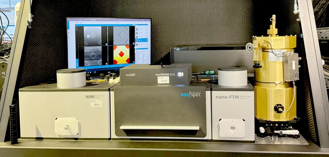
Nanospectroscopy end-station at IRIS Beamline / BESSY II Storage ring
The IR synchrotron light can be used for both imaging and point spectroscopy experiments. Using the broadband synchrotron light the the observed contrast in the recorded images does not contain exact spectral information related to a specific band, but rather represents intensity changes over the whole broad spectrum. The point spectroscopy method is used to reveal the spectral changes at the point of interest. In addition to the synchrotron-based modes, the end station allows to perform pseudo- heterodyne IR imaging using an additional tunable single-frequency laser source.
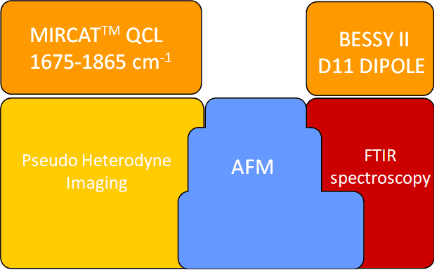
Outline of the nanospectroscopy end-station at IRIS beamline/ BESSY II
Available spectral range
The current configuration of the microscope allows to perform nano-spectroscopy experiments in the spectral range of ~350-3000 cm-1, determined by the available MCT and Si:B detectors, used for the mid- and far-IR spectral ranges, respectively.
Single frequency pseudo- heterodyne IR imaging is a laser source-based modality and is available in the spectral range of 1675-1865 cm-1.

Amplitude of the optical signal collected from the Si-reference sample at the nano-spectroscopy end-station using MCT and Si:B detectors.