Susan Schorr named DGK Chair

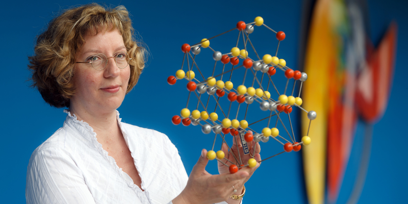
Susan Schorr is Head of the Department of Crystallography.
Prof. Dr. Susan Schorr was elected Chair at the German Crystallographic Society’s (DGK) recent annual conference. The conference took place from March 16-19, 2015, in Göttingen, Germany. Prior to her current appointment, Susan Schorr was head of the DGK’s National Committee.
Susan Schorr is the current head of the Department of Crystallography at the Helmholtz-Zentrum Berlin and is also a professor at the Freie Universität Berlin. The focus of Schorr’s work is on an examination of the underlying structure-property relationships of materials used in energy conversion, for example, in new types of materials for solar cells. In her work, Schorr is drawing on methods for neutron diffraction, synchrotron radiation, and conventional X-rays. In addition, Prof. Schorr is actively engaged in the promotion of junior scientists, as through her work on the faculty of the MatSEC Graduate School and as part of the school’s focus on research management.
DGK, the German Crystallographic Society, is a nonprofit organization whose mission it is to promote crystallography in the educational, research, and industrial settings, as well as in the public sphere. The Society currently has over 1,000 individual members.
www.dgk-home.de
arö
https://www.helmholtz-berlin.de/pubbin/news_seite?nid=14170;sprache=en
- Copy link
-
Battery research with the HZB X-ray microscope
New cathode materials are being developed to further increase the capacity of lithium batteries. Multilayer lithium-rich transition metal oxides (LRTMOs) offer particularly high energy density. However, their capacity decreases with each charging cycle due to structural and chemical changes. Using X-ray methods at BESSY II, teams from several Chinese research institutions have now investigated these changes for the first time with highest precision: at the unique X-ray microscope, they were able to observe morphological and structural developments on the nanometre scale and also clarify chemical changes.
-
BESSY II: New procedure for better thermoplastics
Bio-based thermoplastics are produced from renewable organic materials and can be recycled after use. Their resilience can be improved by blending bio-based thermoplastics with other thermoplastics. However, the interface between the materials in these blends sometimes requires enhancement to achieve optimal properties. A team from the Eindhoven University of Technology in the Netherlands has now investigated at BESSY II how a new process enables thermoplastic blends with a high interfacial strength to be made from two base materials: Images taken at the new nano station of the IRIS beamline showed that nanocrystalline layers form during the process, which increase material performance.
-
Martin Keller elected new president of the Helmholtz Association
The Helmholtz Association has appointed internationally respected US-based scientist Martin Keller as its new president. Her has lived in the United States for nearly three decades, during which he has held various scientific leadership roles at prominent institutions. Since 2015, Keller has directed the National Renewable Energy Laboratory (NREL) in Golden, Colorado. His term begins on 1.11. 2025.
