Department Atomic-Scale Dynamics in Light-Energy Conversion
Ultrafast X-ray and Electron Science
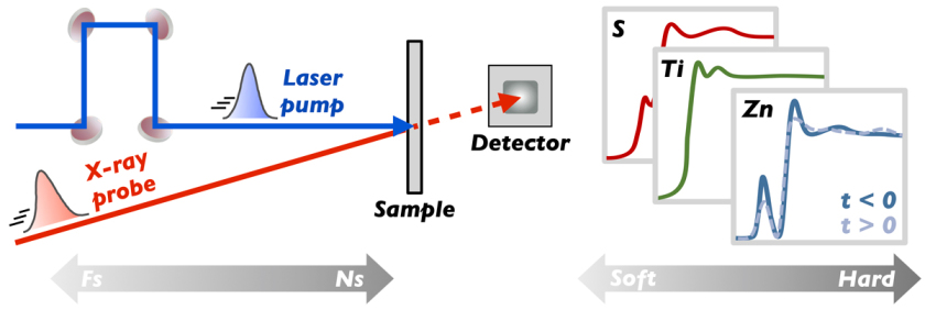
Ultrafast soft and hard X-ray spectroscopy of nanoscale and molecular materials
X-ray absorption spectroscopy (XAS) is particularly appealing for the study of metal-organic and heterogeneous (nano)materials, as it is an element-specific local probe of both the electronic and geometric structure, and it can be implemented in any medium. In our lab, we use short (fs-ps) X-ray pulses from synchrotron radiation facilities or X-ray free-electron lasers (XFEL) to probe the dynamics after excitation with a short laser pulse. We have a particular interest in heterostructured nanomaterials for photo-(electro)catalysis and solar energy conversion, molecular photocatalysts based on first-row transition metals, and strongly cooperative spin-crossover nanomaterials.
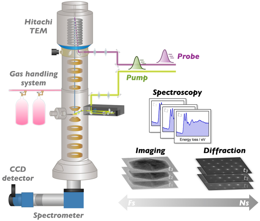
Ultrafast electron microscopy
In collaboration with the CatLab and the group of Prof. Christoph Koch at the Humboldt University we are developing a unique ultrafast electron microscope (UEM). UEM combines the high temporal resolution of ultrafast laser spectroscopy with the superb spatial resolution of transmission electron microscopy (TEM). Compared to ultrafast optical and X-ray methods, UEM exhibits a superior spatial imaging resolution (sub-nm), and it offers the possibility to characterize morphology (via imaging), geometric structure (via diffraction), and electronic structure (via spectroscopy, EELS) of materials - all within the same table-top setup. The UEM apparatus is based on a Hitachi H8110 TEM, which is being modified to enable two pulsed laser beams to enter the ultrahigh vacuum environment of the microscope. One laser beam is used for excitation of the sample with tunable UV/visible wavelengths and pulse durations between ~200 fs and tens of ns. A second UV laser beam is used for the generation of short electron packets by making use of the photoelectric effect at the cathode tip in the microscope gun. By finely tuning the relative time delay between laser excitation (pump) pulses and the electron probing pulses using an optical delay line (fs time scale) or digital delay generators (ns time scale), we are able to make an atomic-scale movie of the photoinduced structural dynamics in individual nanostructures.