IRIS beamline at BESSY II extended with nanomicroscopy

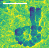
Infrared image of the nucleolus in the nucleus of a fibroblast cell. The scale bar corresponds to 500 nanometres. © HZB
The IRIS infrared beamline at the BESSY II storage ring now offers a fourth option for characterising materials, cells and even molecules on different length scales. The team has extended the IRIS beamline with an end station for nanospectroscopy and nanoimaging that enables spatial resolutions down to below 30 nanometres. The instrument is also available to external user groups.
The infrared beamline IRIS at the BESSY II storage ring is the only infrared beamline in Germany that is also available to external user groups and is therefore in great demand. Dr Ulrich Schade, in charge of the beamline, and his team continue to develop the instruments to enable unique, state-of-the-art experimental techniques in IR spectroscopy.
As part of a recent major upgrade to the beamline, the team, together with the Institute of Chemistry at Humboldt University Berlin, has built an additional infrared near-field microscope.
"With the nanoscope, we can resolve structures smaller than a thousandth of the diameter of a human hair and thus reach the innermost structures of biological systems, catalysts, polymers and quantum materials," says Dr Alexander Veber, who led this extension.
The new nanospectroscopy end station is based on a scanning optical microscope and enables imaging and spectroscopy with infrared light with a spatial resolution of more than 30 nm. To demonstrate the performance of the new end station, Veber analysed individual cellulose microfibrils and imaged cell structures. All end stations are available to national and international user groups.
Funding information: Bundesministerium für Bildung und Forschung [grant No. project 05K19KH1 (SyMS)]; Germany's Excellence Strategy (grant No. EXC 2008-390540038 – UniSysCat).
arö
https://www.helmholtz-berlin.de/pubbin/news_seite?nid=26746;sprache=en
- Copy link
-
Green hydrogen: A cage structured material transforms into a performant catalyst
Clathrates are characterised by a complex cage structure that provides space for guest ions too. Now, for the first time, a team has investigated the suitability of clathrates as catalysts for electrolytic hydrogen production with impressive results: the clathrate sample was even more efficient and robust than currently used nickel-based catalysts. They also found a reason for this enhanced performance. Measurements at BESSY II showed that the clathrates undergo structural changes during the catalytic reaction: the three-dimensional cage structure decays into ultra-thin nanosheets that allow maximum contact with active catalytic centres. The study has been published in the journal ‘Angewandte Chemie’.
-
An elegant method for the detection of single spins using photovoltage
Diamonds with certain optically active defects can be used as highly sensitive sensors or qubits for quantum computers, where the quantum information is stored in the electron spin state of these colour centres. However, the spin states have to be read out optically, which is often experimentally complex. Now, a team at HZB has developed an elegant method using a photo voltage to detect the individual and local spin states of these defects. This could lead to a much more compact design of quantum sensors.
-
Accelerator Physics: First electron beam in SEALab
The SEALab team at HZB has achieved a world first by generating an electron beam from a multi-alkali (Na-K-Sb) photocathode and accelerating it to relativistic energies in a superconducting radiofrequency accelerator (SRF photoinjector). This is a real breakthrough and opens up new options for accelerator physics.
